Mechanism of Action

Alpha1H and the predecessor molecule, HAMLET, are tumoricidal protein-lipid complexes with broad effects against cancer cells of different origins. 

The involvement of specific alpha-lactalbumin peptide motifs in tumor cell death, was examined by synthesizing the N-terminal alpha-helical domain (residues 1-39) or the beta sheet (residues 40-80) domains of human α-lactalbumin (Nadeem et al., 2019, Brisuda et al., 2021). The alpha1 peptide formed complexes with oleic acid (Alpha1H, 1:5) and circular dichroism (CD) spectra detected an increase in alpha-helical structure content in the Alpha1H complex.

Alpha1H triggered a rapid, dose-dependent cell death response, quantified by ATP levels and PrestoBlue fluorescence in human lung (A549), kidney (A498) and murine bladder (MB49) cancer cells. In comparison, the beta-oleate complex lacked tumoricidal activity and tumor cells incubated with individual alpha-helical peptides (35 μM) or oleic acid (175 μM) were not killed. The loss of cell viability was irreversible, as shown after 10 days, using colony assays. It was concluded that the alpha1 peptide forms a tumoricidal complex with oleic acid, which is functionally similar to HAMLET (Brisuda et al., 2021).

HAMLET and Alpha1H initiate the cell death response by integration into the plasma membranes of tumor cells (Storm et al., 2013, Nadeem et al., 2015, Brisuda et al., 2021). The membrane lipid composition differs between healthy differentiated cells and tumor cells and results indicate that membrane lipid conformations serve as surrogate receptors for subsequent signal transduction, leading to tumor cell death (Nadeem et al., 2015, Ho et al., 2022). Cell membrane responses were observed via rapid Na+ and K+ fluxes and pretreatment of the cells with Na+ or K+ flux inhibitors reduced cell death by 40-50%, linking the membrane response and ion flux to tumor cell death (Figure 1).

Finally, the Alpha1H complex (labeled with AlexaFluor568) is rapidly internalized by A549 tumor cells and translocates to the nuclei, where it triggers double-strand DNA breaks as measured by TUNEL staining. The cells die by an apoptosis-like mechanism (Figure 1).

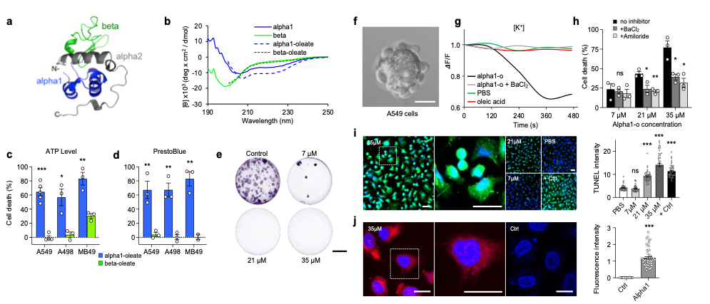
 

Figure 1: Mechanism of Action of Alpha1H

Figure 1 legend: a. Representation of α-lactalbumin protein structure, indicating the alpha1 (blue), beta (green) and alpha2 (gray) domains. b. Evidence of alpha1 peptide and oleic acid interaction by CD spectroscopy, forming the Alpha1H complex. c,d. Evidence of short-term cell death response to Alpha1H, in human lung (A549), kidney (A498) and murine bladder (MB49) cancer cells. e. Evidence of long-term effect of Alpha1H on cell death. f. Evidence of membrane perturbations by Alpha1H on cancer cells. g,h. Evidence of ion flux response to Alpha1H treatment and inhibition of Alpha1H-induced cell death by ion flux inhibitors. i. Evidence of DNA strand breaks in Alpha1H-treated cells. j. Evidence of Alpha1H (red) internalization in treated cells (blue nuclei).

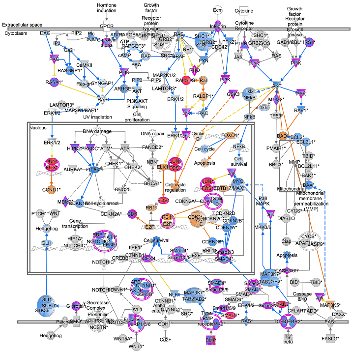
Classical oncogenes like MYC, RAS and HIF1a were further identified as determinants of HAMLET sensitivity in a short hairpin (sh) RNA screen of cancer cells, confirming that cells targeted by HAMLET fulfill generally accepted criteria. Oncogene signaling pathways are inhibited by Alpha1H in cells and tissues exposed to the complex and the expression of cancer related genes is markedly reduced (Storm et al., 2011; Nadeem et al., 2019; Brisuda et al., 2021; Tran et al, 2023).

The inhibition of cancer-related gene expression in treated lung carcinoma cells is shown in Figure 2. Alpha1H targeted and inhibited cancer-related genes in cancer cells.

Figure 2: Inhibition of Cancer-Related Transcriptional Networks

– Molecular mechanism of action

Figure 2 legend: Gene expression analysis of RNA isolated from lung carcinoma cells exposed to Alpha1H (unlabeled, 35 µM) or PBS for 1 hour. Molecular mechanisms of cancer genes were inhibited after 1 hour, including Ras oncogene signaling, PI3kinase, which regulates transcription, TGFbeta and osteopontin, which are cancer related growth factors. Further effects on gene expression included cell cycle progression, NOTCH and EP300 signaling.

These results identify alpha1H as a potent regulator of gene expression in cancer cells, translated into a suppressive effect on the expression of cancer-associated genes and gene networks.