The History of HAMLET

Since 1995, the Svanborg laboratory has also been pursuing the discovery of HAMLET; a human milk complex with broad tumoricidal activity. 

HAMLET is the first member in a new and expanding family of lipid-bound partially unfolded proteins with tumoricidal activity and offers an interesting new tool in tumor biology. Furthermore, HAMLET has therapeutic activity against experimental and human tumors. In a placebo-controlled clinical study, topical administration of HAMLET removed skin papillomas, without side effects. In patients with bladder cancer, HAMLET triggered rapid shedding of tumor cells into the urine and caused a reduction in tumor size. Recent work has shown that the broad sensitivity of tumor cells to HAMLET relies on the oncogene repertoire, especially on c-Myc expression levels. Furthermore, the tumor killing effect of HAMLET reflects the metabolic state of the tumors, defined by specific glycolytic enzymes. Ongoing studies focus on the ‘’switch’’ turned on by HAMLET in tumor cells, to initiate the death response. Therapeutic models include colon cancer, with promising results. One of the main goals is to elucidate the structural basis of HAMLET and the difference is HAMLET sensitivity between tumor cells and healthy cells.

HAMLET, is a human molecule that kills cancer cells. we have characterised the molecule and its mechanism of action and have shown in clinical trials that HAMLET works in patients.

HAMLET is a new drug candidate with astonishing but well-documented properties.

  1. HAMLET kills a broad range of tumor cells but spares healthy, mature cells (Proc. Natl. Acad. Sci, 1995 and 2000).
  2. HAMLET kills more than 40 different types of tumour cells, including those most difficult to treat with available drugs.
  3. HAMLET kills tumour cells by a natural non-toxic mechanism. Thus, unlike most current cancer drugs, HAMLET does not appear to damage healthy tissues.
  4. HAMLET occurs naturally in human milk, and may contribute to the lowered cancer incidence in breast-feeding mothers and their children.
  5. HAMLET can be produced in large quantities at drug quality.

We will develop the concept of innate immunotherapy in cancer, using HAMLET as a model. Studies of structure and mechanism of action will be combined with proof of concept studies of therapeutic efficacy in animal models and patients. 

HAMLET, a new concept for cancer therapy

Background

Few molecules destroy cancer cells without harming healthy tissues. Instead, the side effects of current cancer drugs have become an additional therapeutic area. As a result of new technologies and conceptual approaches, more targeted cancer therapies are starting to appear but the lack of specificity for tumor cells remains a significant problem. Identifying how tumor cells differ from healthy cells is therefore a great challenge, especially mechanisms of more tumor specific cell death as a basis for more tumor selective therapies.

New concepts and innovative approaches are needed to achieve tumor specific cell death and to develop more tumor selective therapies. Increasingly, biotherapies are showing promise as anticancer agents and are still a relatively untapped source of new molecules with novel mechanisms of action.

HAMLET (Human -lactalbumin Made LEthal to Tumor cells) is a complex of partially unfolded α-lactalbumin and oleic acid that kills tumor cells and immature cells but not fully differentiated healthy cells. 

We discovered the activity of HAMLET, while using human milk fractions to block bacterial adherence to lung carcinoma cells. One milk fraction killed the tumor cells and the molecular complex responsible for this effect was identified as a folding variant of α-lactalbumin bound to oleic acid. HAMLET is the first member in a new class of potent tumoricidal molecules is formed by self-assembly of milk proteins and oleic acid.

The human variant HAMLET kills a wide range of tumor cells in vitro and shows broad but

 selective therapeutic efficacy in patients and cancer models. Its efficacy as a selective killer of tumor cells has been documented in vitro and in vivo in several animal models, including human brain tumor xenografts in nude rats, murine bladder cancer and colon cancer in the APCMin+/- mice. In clinical studies, purified HAMLET from human breast milk has shown therapeutic efficacy against skin papillomas and dramatic effects in bladder cancer patients.

Since the discovery of HAMLET in our laboratory, we have characterized the structure of the HAMLET complex, its cellular targets and therapeutic efficacy in animal models and clinical studies.

1. HAMLET triggers a broad tumoricidal response with apoptosis-like features

The early collaborative studies focused on the apoptotic response that accompanies death in HAMLET treated tumor cells and the role of mitochondria in this process. The morphologic changes in dying tumor cells resembled apoptosis, with nuclear condensation, loss of cytoplasm and membrane blebbing. Using the DNA fragmentation technology of the Orrenius laboratory, HAMLET was shown to trigger typical DNA laddering. Tumor cells from different tissues shared the apoptotic features but normal cells in primary culture were resistant, suggesting an unexpected degree of tumor specificity. Compared to classical apoptosis inducers like Etoposide, the morphological changes and loss of viability were more rapid and more tumor specific.

In experiments with the Orrenius laboratory, we observed swelling and depolarization of mitochondrial membranes, accompanied by the release of Cytochrome C, activation of caspase 3, caspase 9 and phosphatidylserine exposure on the outer leaflet of the plasma membrane. These findings suggested that the cells were dying by apoptosis, initiated at the level of the mitochondria. Paradoxically, tumor cells were not rescued by the pan-caspase inhibitor ZVAD, pointing to an alternative death mechanism. In later studies, we further showed that HAMLET-induced cell death is independent of the host BCL-2 and p53 genotype. Overexpression of the anti-apoptotic proteins, BCL-2 and BCL-XL did not significantly alter the cell death response and a Tet-inducible p53 expression vector had no effect. Parallel work on the effects of HAMLET on bacteria cell death have shown that such morphological changes and biochemical responses can also be seen in prokaryotes.

2. Cellular and molecular targets

2.1. Oncogenes determine the sensitivity to HAMLET

HAMLET kills a large number of different tumor cells, in vitro, suggesting that the complex targets conserved mechanisms of cell death. In collaboration with Cold Spring Harbor Laboratory, we identified classical oncogenes like MYC, RAS and HIF1α as genetic determinants of HAMLET sensitivity in an shRNA screen, suggesting that cells targeted by HAMLET fulfill generally accepted tumor criteria. Still, the mechanism of action of HAMLET is unusual, as the complex interacts with a number of molecular targets and cellular compartments. After initial membrane interactions, HAMLET is internalized by the tumor cells, interacts with lysosomes, mitochondria and proteasomes and translocates to the nuclei. Organelle-specific molecular interactions also lead to proteasome inhibition, chromatin modification, transcriptional inhibition and death

Cellular and molecular targets

2.2. Membrane perturbations and ion fluxes

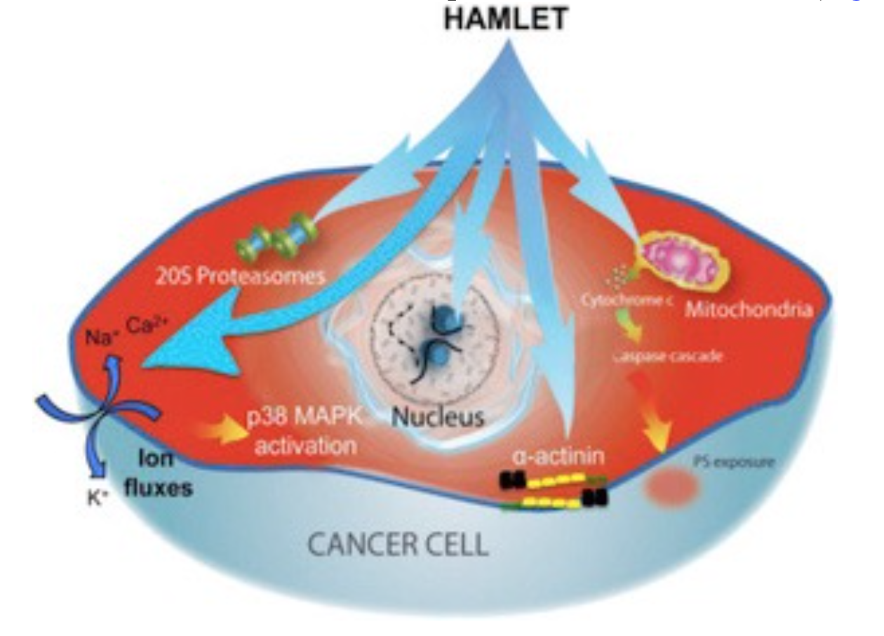
HAMLET integrates into the membranes of tumor cells and triggers rapid ion fluxes, which activate downstream signaling pathways involved in cell death. Three critical features of the membrane response to HAMLET have been identified. (1) HAMLET interacts with lipid membranes in a protein receptor independent manner, as shown by the insertion of HAMLET into the membranes of giant unilamellar vesicles, disruption of the spherical shape and rapid tubulation, leading to a reduction in lumen size. (2) HAMLET creates a new membrane compartment for interactions with cellular targets and inhibits critical Ras oncogene driven signaling pathways. (3) This response to HAMLET does not occur in primary non-transformed cells, indicating tumor selectivity. The results suggest that HAMLET produces membrane conformations that serve as surrogate receptors for subsequent signal transduction, leading to tumor cell death. These effects are specific to the HAMLET complex, as the individual components of the complex, native alpha lactalbumin and oleic acid, did not affect the integrity of model membranes. The results indicate that both the partially unfolded protein and oleic acid are required to alter the structure of tumor cell membranes.

Ion fluxes regulate cellular homeostasis, through a multitude of effector functions. In parallel with the membrane changes, HAMLET activates rapid ion fluxes, with an influx of Na+ and Ca2+ and an efflux of K+ ions, as shown by real time confocal imaging, patch-clamping and fluorometry. The Na+ and K+ fluxes are thought to drive the cell death response, as ion flux inhibitors prevent HAMLET internalization, transcriptional responses and tumor cell death. These include amiloride, which inhibits the Na+/H+exchange and barium chloride (BaCl2), which inhibits K+ fluxes. HAMLET has not been shown to activate preformed ion channels, suggesting that channel-independent membrane permeabilization mechanisms might be involved.

Further mechanistic insights into this process have been gained, using synthetic alpha-helical peptides. Peptides covering the alpha1 and alpha2 domains of the protein triggered rapid ion fluxes in the presence of oleate and were internalized by tumor cells, causing rapid and sustained changes in cell morphology. The alpha peptide-oleate bound forms also triggered tumor cell death with comparable efficiency as HAMLET. In addition, shorter peptides corresponding to those domains were biologically active.

The results identify HAMLET as a membrane-perturbing agonist that triggers lethal ion fluxes in tumor cells.

2.3. Effects on nucleotide-binding proteins – kinases and GTPases

The ion fluxes activate a rapid p38 MAPK response as shown by transcriptomic analysis of HAMLET-treated tumor cells. In parallel, ERK1/2 phosphorylation is inhibited, consistent with the shift from proliferation to cell death. The activation of p38 and inhibition of ERK1/2 phosphorylation was reversed by ion flux inhibitors (amiloride or BaCl2), confirming the ion flux dependence. Importantly, pharmacological inhibitors of p38α and p38β delayed tumor cell death, as did p38-specific siRNAs. In contrast to tumor cells, normal differentiated cells showed weaker ion flux responses and less prominent changes in global transcription.

These findings suggested that affinity for conserved molecular motifs explains the apparent multitude of HAMLET targets in tumor cells. To identify such targets, we performed a proteomic screen of 8000 human proteins. By direct binding, we identified a large number of nucleotide binding proteins as HAMLET targets. These included 3 ATPases, 24 members of the Ras family of GTPases and 111 Kinases representing all branches of the kinome tree. In a kinase activity array, HAMLET acted as a pan-kinase inhibitor, reducing the activity of about 69% of the 476 kinases tested. This broad kinase inhibition was confirmed in protein lysates from HAMLET treated lung carcinoma cells, using an antibody microarray detecting phosphorylated proteins. Furthermore HAMLET was shown to co-localize with the Ras family of GTPases in membrane clusters and Ras activity was inhibited. The results identify HAMLET as an inhibitor of kinases and GTPases, to which tumor cells are addicted.

The results identify HAMLET as a potent and broad kinase inhibitor with specificity for tumor cells.

2.4. Chromatin interactions

In early studies our group has shown that HAMLET crosses the cytoplasmic membrane and accumulates in tumor cell nuclei. The accumulation of HAMLET in tumor cell nuclei was initially demonstrated by confocal microscopy of biotinylated HAMLET and fluorophore-labeled streptavidin and was confirmed using radiolabeled HAMLET. By far western blot and mass spectrometry analysis (MALDI-TOF), histones H2B, H3 or H4, were identified as nuclear HAMLET targets and HAMLET was shown to interfere with the formation of nucleosomes in the salt-jump assay. Furthermore, the sensitivity of tumor cells to HAMET is affected by chromatin acetylation, as histone deacetylase inhibitors open up the chromatin to HAMLET and synergistically kill tumor cells.

The results suggest that HAMLET may ‘seal the fate’ of dying tumor cells, through high affinity histone interactions and perturbation of the chromatin structure.

2.5. Proteasome inhibition

Proteasomes are crucial for extra-lysosomal protein degradation of endogenous misfolded proteins taking place in the barrel shaped 20S proteasome core. In a proteomic screen, catalytic proteasome subunits were identified as HAMLET targets. Furthermore, as α-lactalbumin is partially unfolded in the HAMLET complex, we hypothesized that the complex is targeted to the 20S proteasome for degradation. We found that HAMLET co-localizes with proteasomes in tumor cell cytoplasm and nuclei and detected an inhibitory effect of proteasome activity in whole cell extracts from HAMLET treated tumor cells. Interestingly, we also obtained structural evidence for proteasome disintegration by HAMLET, after incubation of HAMLET with intact 20S proteasomes, in vitro. Based on the above observations, we concluded that inhibition of proteasome activity might contribute to HAMLET-induced tumor cell death.

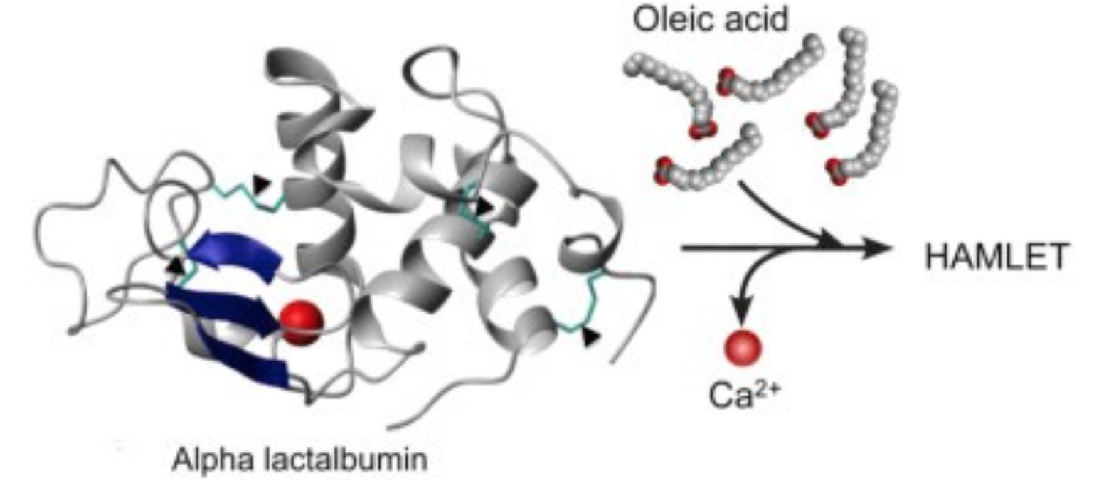
3. Structure of HAMLET complex

HAMLET is the first member in a new class of tumoricidal protein-lipid complexes, formed by partially unfolded α-lactalbumin and oleic acid. α-Lactalbumin is the most abundant protein in human breast milk and the tightly packed globular conformation is stabilized by four disulfide bridges and a divalent calcium ion, with C- and N-terminal α-helical domains separated by a β-sheet domain.

The native, globular state is defined by high affinity interactions with a strongly bound calcium ion (Ca2+), and conditions that release Ca2+, such as low pH or EDTA treatment are accompanied by a loss of tertiary structure definition. As a result the protein adopts a stable intermediate fold and forms a molten globule with loss of tertiary structure and retained secondary structure as demonstrated by near- and far-UV CD. In addition, ANS fluorescence is increased, reflecting the exposure of hydrophobic domains. Differences in surface topology have also been detected by limited proteolysis, compared to the native protein. In HAMLET, α-lactalbumin retains its partially unfolded characteristics even at physiological solvent conditions, suggesting that the binding of oleic acid stabilizes the protein in the partially unfolded state.

Moreover, HAMLET tolerates a certain degree of sequence variation, as purified α-lactalbumins from different species formed tumoricidal complexes with the fatty acid cofactor, oleic acid. This includes bovine, equine, caprine and porcine α-lactalbumins. The conversion yield for α-lactalbumin derived from other species is lower than for the human protein, however. 

Even though these proteins can form HAMLET-like complexes in vitro after purification and addition of the lipid, HAMLET-like complexes are not formed by low pH treatment of milk from other species. HAMLET formation has so far only been detected in human milk, reflecting both the α-lactalbumin structure and the fatty acid composition.

The low-resolution structure of HAMLET has recently been solved using small angle X-ray scattering (SAXS). The SAXS structure shows a two-domain conformation with a large globular domain and an extended C-terminal domain (Fig. 3). According to the SAXS structure, HAMLET exists as a monomer in solution with a molecular mass of 15 ± 2 kDa.

Unfolding of α-lactalbumin is not sufficient to achieve cytotoxicity. Mutant proteins that fail to fold to the native state are not cytotoxic for tumor cells. Tested protein variants include the high-affinity Ca2+ binding site mutant (D87A) and the fully reduced cysteine-free mutant (rHLA All-Ala), in which all cysteine residues were substituted for alanines. The mutant proteins became cytotoxic after addition of the lipid cofactor, however. The lipid alone is significantly less cytotoxic than the HAMLET complex, at concentrations comparable to those present in the HAMLET complex.

3.1 Gain of function by loss of tertiary structure definition

This gain of tumoricidal activity by loss of 3D structural definition may appear paradoxical. The ‘one gene – one protein – one function’ paradigm considers the native state of a protein as the functional conformation, which is usually equated with the lowest free energy state of a given molecule. HAMLET is the first example of a protein that exhibits a well-defined function in its native state and then acquires a new and beneficial function after partial unfolding. Our findings suggest that a change in fold, in response to changing tissue environments, may allow a single polypeptide chain to exert vastly different, beneficial biological functions in different tissue compartments.

Complexes similar to HAMLET, which consist of protein and fatty acid constituents, exhibit cytotoxic activities. Examples of such complexes include BAMLET (Bovine Alpha-lactalbumin Made LEthal to Tumor cells), ELOA (Equine Lysozyme with Oleic Acid), and other oleic acid complexes with camel α-lactalbumin, β-lactoglobulin or pike parvalbumin. These findings suggest that the ability to form lipoprotein complexes may be a general feature of partially unfolded proteins.

4. Therapeutic and prophylactic effects of HAMLET

HAMLET has shown therapeutic efficacy in three cancer models; colon cancer, bladder cancer and a human glioblastoma xenograft model. In addition, HAMLET has been tested in a placebo-controlled study of human skin papillomas and in patients with bladder cancer. In each of these cases, we observed positive and interesting effects.

4.1. Human studies

a) Skin papillomas. We demonstrated therapeutic efficacy of HAMLET against human skin papillomas, in a placebo-controlled, blinded clinical study [44]. Patients with severe, therapy resistant papillomas on hands and feet received HAMLET or saline solution daily for 3 weeks. Topical application of HAMLET reduced the lesion volume by more than 75%. Moreover, a significant decrease in lesion volume was observed in all HAMLET treated patients and complete resolution of all lesions had occurred in about 83% of the HAMLET-treated patients after two years.

b) Bladder cancer. To examine if bladder cancer cells respond to HAMLET, patients with superficial bladder cancer received injections of HAMLET locally, into the bladder, on five consecutive days preceding bladder surgery. A rapid response was detected, with excretion of large numbers of tumor cells into the urine after two hours. Most of the cells were dead and showed evidence of apoptosis. By cystoscopy, a reduction in tumor size was detected at the time of surgery and apoptotic cells were seen in biopsy specimens.

4.2.  Animal models

a) Bladder cancer. Local instillation of HAMLET caused a reduction in tumor development, in mice with bladder cancer. Whole body fluorescence imaging showed that HAMLET is retained in tumor bearing mice compared to tumor free mice. Furthermore, five intra-vesical HAMLET instillations decreased tumor size and significantly delayed tumor development in tumor bearing mice compared to controls that received α-lactalbumin or phosphate buffer.

b) Glioblastomas. Therapeutic efficacy of HAMLET against human glioblastomas was investigated in ratsnu/nu after xenotransplantation of human glioblastoma cells. Local infusions of HAMLET delayed tumor development and prolonged survival. HAMLET penetrated throughout the tumor and triggered apoptosis in tumor cells. Importantly, there was no evidence of HAMLET toxicity for the normal brain.

c) Intestinal cancer. The therapeutic efficacy of HAMLET was investigated in a model of human colon cancer. APCMin/+ mice develop intestinal tumors that serve as a model of human disease. APC mutations occur in the majority of patients with colon cancer and in families with inherited susceptibility to colon cancer. Peroral administration of HAMLET caused a significant reduction in tumor size and polyp number in this model. In addition, HAMLET accumulated specifically in tumor tissue. In parallel, the expression of key oncoproteins was reduced after HAMLET administration, including β-catenin, Ki67, COX2 and VEGF. By whole genome transcriptomic analysis HAMLET was shown to inhibit the expression of genes in the Wnt signaling pathway in APCMin/+ mice. Furthermore, we supplied HAMLET into the drinking water of young mice for ten weeks, from the time of weaning. This treatment reduced tumor development by 60%, suggesting that HAMLET acts prophylactically.

In conclusion, these studies have established that local HAMLET administration is effective, with therapeutic and prophylactic effects against several different tumors. Importantly, we did not observe toxic effects on healthy tissues in treated patients or animals.